Technology best of its kind on campus

By Karyn Houston
Plant & Microbial Biology
A microscopy expert at UC Berkeley has won a grant to purchase an amazingly powerful new microscope that will enable scientists to study the tiniest of organisms.
"This new microscope will enable researchers to see objects that are impossible to see using technology available at Berkeley today," said Steve Ruzin, director of the Biological Imaging Facility, located on the UC Berkeley campus in Koshland Hall, in the College of Natural Resources.
Ruzin is an expert in microscopes and his cutting-edge Biological Imaging Facilty serves thousands of faculty, students and staff. Not only are a wide variety of microscopes available for researchers to use, but the lab also teaches students and other researchers about microscopy, the technical field of using microscopes to view samples and objects that cannot be seen with the naked eye.
"Cells have bustling shipping centers," said Amita Gorur, a graduate student in the Randy Schekman Lab at UC Berkeley. "The Elyra PS.1 Super Resolution microscope will allow me to track and visualize cargo on a freight car moving along cellular rail roads from destination A to B in real time. These cellular shipping units are so small that only the resolution achieved by this microscope will allow us to see them. That's powerful!"
Differentiating Objects from One Another

The new $600,000 instrument, purchased with a National Institutes of Health grant, is a "Structured Illumination Microscope" that allows researchers to image and differentiate different parts of a cell, using different fluorescent dyes.
"In microscopy, and any optical system, to 'see' something is the ability of the system (microscope, telescope, eye) to determine whether two closely spaced objects are, in fact, two objects or really only one larger object," Ruzin said. This "limit of resolution” is determined by the wavelength of light that is used to illuminate the sample. The resolution limit was defined in the 1880s by the German scientist Ernst Abbe, and it is still valid today.
"In practical terms this means that the smallest object that can be resolved in a light microscope is about one third of a micrometer, or 300 nanometers," Ruzin said. To put that size into perspective:
o The period at the end of this sentence is about a millimeter in diameter.
o A single bacterium is 1,000 times smaller than a period, or about 1 micrometer.
New Technology
In the last few years three separate technologies have become available that overcome the limit of resolution. The new microscope uses one of these technologies called “Structured Illumination," that illuminates the sample with a known pattern of light.
The illumination pattern induces a complex light pattern that is emitted from the sample. Subsequent computer processing of the emitted pattern reveals sub-resolution structures within the sample. The new microscope will achieve a resolution of 100nm, and will be able to see objects that are 10 times smaller than a bacterium, or 10,000 times smaller than a period.
 This ability to resolve objects smaller than the theoretical limit is the reason a microscope like this is called a “Super-Resolution Microscope”. It will enable researchers to see:
o the arrangement of proteins within bacteria
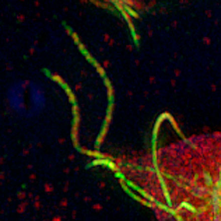
o the micro-structure of Giardia flagella
o the sub-resolution arrangement of proteins on chromosomes
o the presence and arrangement of magnetic particles inside bacteria
Arash Komeili’s lab in the Department of Plant & Microbial Biology is one of the many labs on campus that will benefit from the new instrument.
The Komeili group studies magnetosomes, bacterial organelles 50-70 nm in diameter that control the formation of magnetic nanoparticles. Due to their small size and tight arrangement within the cell, conventional fluorescence microscopy cannot distinguish between individual magnetosomes.
“We anticipate that the improved resolution of the Structured Illumination Microscope will allow us to study the dynamics of specific proteins at the individual magnetosome level,” Komeili said.
Important Links
CNR Biological Imaging Facility Website: microscopy.berkeley.edu
